Enjoy a More Proactive, Personalized Approach to Your Skin Health
You deserve healthy, glowing skin — and a customized treatment plan that meets the specific needs of your body.
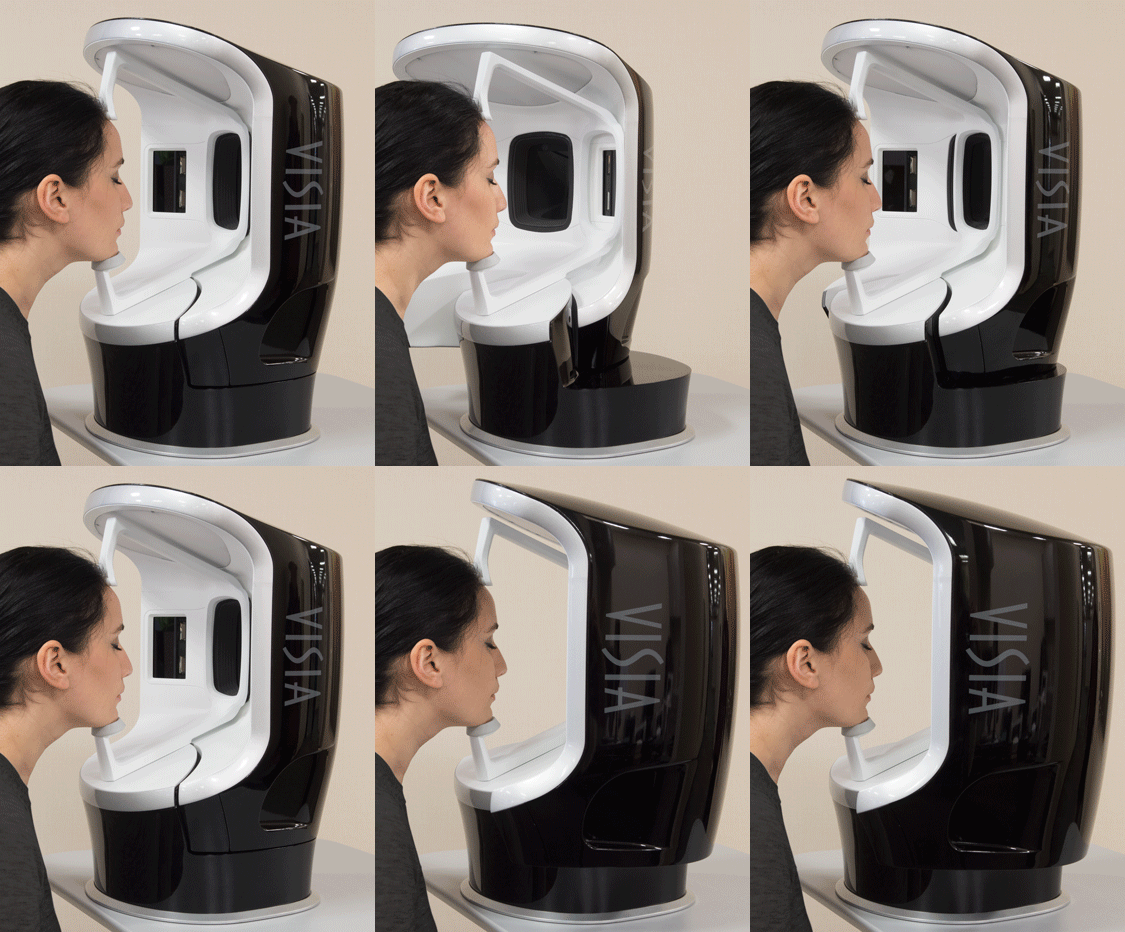
At Chicago Skin Clinic, we offer VISIA Skin Analysis, a state-of-the-art imaging system that helps us get a deeper understanding of your skin’s unique characteristics.

With VISIA, Dr. Danilo Del Campo and Dr. Danny Del Campo can accurately assess your skin’s health, identify any problem areas, and develop a customized treatment plan tailored specifically to your skin’s needs.
So say goodbye to wrinkles, dark spots, and acne — and hello to the clear, youthful-looking skin you’ve always wanted.
What Is VISIA Skin Analysis?
The VISIA Complexion Analysis system is a state-of-the-art technology that helps reveal damage and signs of aging both on and beneath your skin which may otherwise be undetectable by the human eye.
What Does VISIA Skin Analysis Mean for Your Skin Health?
VISIA Skin Analysis at Chicago Skin Clinic help us to diagnose your unique skin conditions earlier and more accurately — so you get more proactive, personalized treatments tailored specifically to your unique skin conditions.
Here’s how: Using this comprehensive digital scan analysis, Dr. Danilo Del Campo and Dr. Danny Del Campo can analyze surface-level skin conditions — including acne spots and blemishes — as well as deep, underlying injuries such as ultraviolet damage and pigmentation.
Additionally, VISIA can help treat signs of aging closer to the source. By more accurately revealing the presence of expressive and static lines and wrinkles, as well as any redness or build-up of bacteria (porphyrins) within the skin, we can maximize the repair and correction of your skin health.
How Does VISIA Skin Analysis Work?
The VISIA Skin Analysis System works by using cross-polarized and UV lighting to take multiple images of your face from three precise angles.
Using these thorough, multi-spectral images, Dr. Danilo Del Campo and Dr. Danny Del Campo can record more measurements of the surface and subsurface of your skin. This not only helps you see what’s going on with your skin right now, but it can also help you more accurately review how your current treatment results are progressing.
A printed report and a personalized online link, which you can view at home, provide precise details of your imaging session. This comprehensive information helps our board-certified dermatologists determine the best treatments and plan of care to meet your skin needs. The thorough images also help to review the results of your current rejuvenation and skin care treatment to decide whether we need to recommend any changes.
What Eight Key Areas Are Analyzed?
1. Spots
Typically brown or red skin lesions — including freckles, acne scars, hyperpigmentation, and vascular lesions — spots are distinguishable by their distinct color and contrast from the background skin tone.
2. Pores
These are circular surface openings of sweat gland ducts. Due to shadowing, pores appear darker than the surrounding skin tone and are identified by their darker color and circular shape.
3. Wrinkles
These furrows, folds, or creases in the skin increase in occurrence due to sun exposure and are associated with decreasing skin elasticity.
4. Texture
Texture measures skin color and analyzes skin smoothness by identifying gradations in color from the surrounding skin tone, as well as peaks and valleys on the skin surface that indicate variations in the surface texture.
5. Porphyrins
These bacterial excretions can become lodged in pores and lead to acne. Porphyrins fluoresce in UV light and exhibit circular white spot characteristics.
6. UV Spots
These spots occur when melanin coagulates below the skin surface due to sun damage. UV spots are generally invisible under normal lighting conditions. The selective absorption of the UV light by the epidermal melanin enhances its display and detection by VISIA.
7. Red Areas
These colorful areas can present a potential variety of conditions, such as acne, inflammation, Rosacea, or spider veins.
RBX Technology in VISIA detects blood vessels and hemoglobin contained in the papillary dermis — a sub-layer of skin — that give these structures their red color.
Acne spots and inflammation vary in size but are generally round. Rosacea is usually larger and diffuse compared to acne, and spider veins usually are short and thin and can be interconnected in a dense network.
8. Brown Spots
Brown spots are lesions on the skin such as hyperpigmentation, freckles, lentigines, and melasma. They occur as a result of an excess of melanin, which is produced by melanocytes found in the bottom layer of the epidermis.
Get Proactive About Your Face & Skin
By learning more about your skin, you can better protect your body’s biggest organ from damage caused by the sun, aging, or undiagnosed conditions.
Schedule a consultation with Dr. Danny Del Campo or Dr. Danilo Del Campo at our Chicago office. We can incorporate the VISIA Skin Analysis into your cosmetic visit so both you and Chicago Skin Clinic can get a more complete picture of the current condition of your skin.
 SCHEDULE NOW